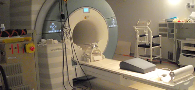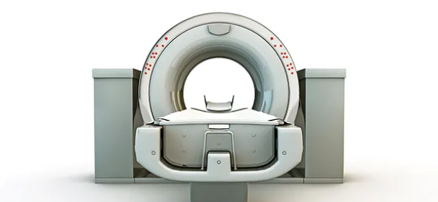




Today, virtually all medical images are available in digital form. Such an image may be a simple 2D representation (digital radiography), a spatial 3D representation (a set of 2D images of a volume, such as an MRI scan of a brain), a 2D + time representation (showing how the perfusion of a contrast agent through an organ changes over time), or even an n-dimensional representation, when the image pixels are described by n values (multispectral imaging). In every case, a medical image is therefore simply an n-dimensional array (or matrix). Each entry in this array is called a pixel in 2D or a voxel in 3D.
Mathematics, naturally
--------------------------------
Although physicians rely heavily on their experience and knowledge to interpret the information carried by these images, the volume and quality of the images obtained make it impossible to use the data to their full potential. Visualization remains two-dimensional—on a screen or photographic film—and is sometimes incomplete, while human expertise allows few, if any, quantitative assessments, even though these are essential for properly analyzing the clinical problem under investigation. Representing images as matrices naturally suggests using mathematical tools to process medical images. This draws on a wide range of mathematics, from geometry and linear algebra to statistics, probability, and partial differential equations.
The tools developed are used in image acquisition as well as in modeling, processing, and information extraction. Software routinely used in clinical practice to manipulate these images also requires the development of suitable visualization tools.