



Tangente: What do mathematics and computer science contribute to your clinical work in brain imaging? What do they contribute to your research?**
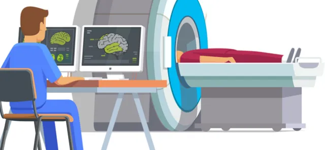
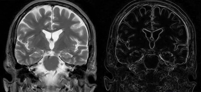
C.H.: First of all, brain imaging would not exist without physics and, a fortiori, without mathematics. Brain imaging encompasses anatomical imaging, which describes the structure of the brain; flow imaging, for blood or cerebrospinal fluid; metabolic imaging, which examines biochemical components through spectroscopy; and functional imaging, which produces maps of brain activation.
The physiology of the brain must therefore be modelled and formalized, since some of its properties are used to create a signal that the machine can detect. Examples include differences in the absorption of X-rays by tissue in computed tomography, proton-spin relaxation in MRI and radioactive emissions in nuclear medicine. We must then model how the physical signal recorded in this way is transformed into an image that a clinician can interpret. And this chain of computations, leading from biological tissue to its image, requires powerful computers!
Modelling and statistical tests
----------------------------------
Furthermore, functional imaging, which seeks to identify the regions of the brain activated during specific mental tasks, requires even more sophisticated mathematical processing. This includes, for example, modelling the brain’s spontaneous response or its response to experimental stimuli, as well as statistical tests to ensure that the results are significant. These brain activation maps take the form of directed graphs, with functionally connected brain regions involved in a particular mental task as nodes and causal relationships between them as directed links; they vary over time.