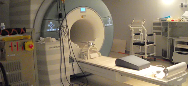




How large is this kidney tumor? Where is the small structure in the brain that I want to target during surgery? Is the stent I placed in the artery positioned correctly? To answer these and many other questions, doctors need a way to locate an organ or object, measure it, or quantify its activity. The underlying problems belong to image segmentation, one of the most interesting and challenging problems in computer vision.
Identifying regions
-------------------
Segmentation consists of identifying and defining the objects present in an image: the image is no longer a collection of juxtaposed pixels (in 2D) or voxels (in 3D), but a collection of disjoint regions. Ideally, these regions have a meaning—an object, structure, signal, or background—beyond their own visual characteristics, such as grayscale values or shape. Methods for identifying regions in an image fall into two groups:
• The region-based approach: pixels sharing a common characteristic, derived for example from their intensity, are grouped together; the criterion is evaluated either in absolute terms (intensity values) or relatively (intensity differences); decision rules are then specified in the form of thresholds defining ranges of values; pixels whose values lie between the thresholds and that are connected to one another (known as connected pixels) form a single region;
• the boundary-based approach: boundaries are placed where the local variation of a criterion reaches a maximum; a boundary is a closed curve, or a closed surface in a 3D image, one pixel thick.