



Radiography, the first medical imaging technique (see above), is the most widely used method for examining the skeleton. To visualize blood vessels, doctors perform angiography. This requires the injection of a contrast agent (usually iodine), which absorbs X-rays.
Scintigraphy is a nuclear-medicine technique: it uses radioactivity for imaging and for treating patients. Patients are given a radioactive tracer that binds to an organ or whose components are carried along by a flow. The tracer emits photons, which a camera detects. This makes it possible to track flows or observe an organ's function in real time.
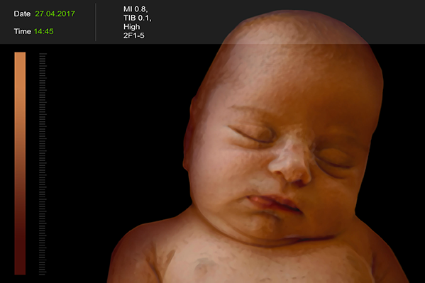
Ultrasound is used in ultrasound imaging. The echo reflected by the organ is used to reconstruct an image in real time. Easy to use, ultrasound is increasingly being used for 4D imaging (3D representations of organs in motion), mainly to visualize heart motion and fetuses. As with MRI, neither a radioactive tracer nor a contrast agent needs to be injected into the patient.
The first radiograph
------------------------